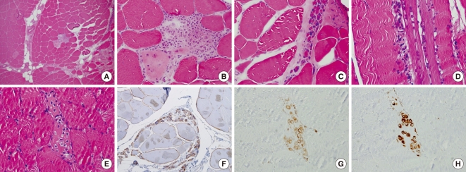
Fig. 1.
Muscle biopsy specimen of right vastus lateralis muscle. It shows variable sized muscle fiber, fatty infiltration, perimysial fibrosis, focal inflammatory infiltrates with the suspicious area of perifascicular atrophy in low power (A) and high power view (B). Perimysial atrophy mimicking dermatomyositis are also shown (C). Also regenerating fibers with inflammatory infiltration was shown (D, E). Dystrophin staining showed variable different staining pattern from normal to interruped mosaic pattern (F). Inflammatory cells are mainly macrophage-lineages on LCA (G), CD 68 (H) special staining with rarety of CD 4 and CD 8 positive cells (not shown).

