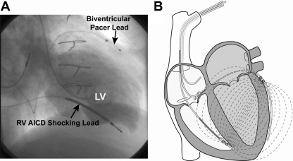
Fig. 1.
A: biplane left ventriculogram from a patient with congestive heart failure and a previously implanted automatic implantable cardiac defibrillator (AICD)/biventricular pacer, demonstrating how the leads span the left ventricle (LV) blood from the right ventricle (RV) apical septum to the lateral LV epicardium. B: the admittance electric field lines from the RV apical septum to the lateral LV epicardium.

