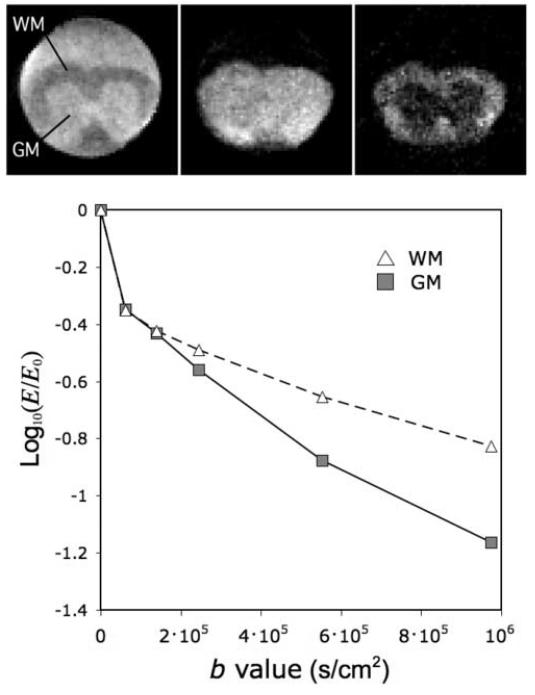
Figure 8.
Diffusion-weighted images of a fixed mouse spinal cord acquired using the Micro-Z with diffusion gradients perpendicular to the cord axis and b = 0.6, 2.5, 9.7 ×10⁵ s/cm2 (left to right). The graph shows different rates of signal loss as a cause of WM/GM contrast inversion at high b-value.

