
Figure 4.
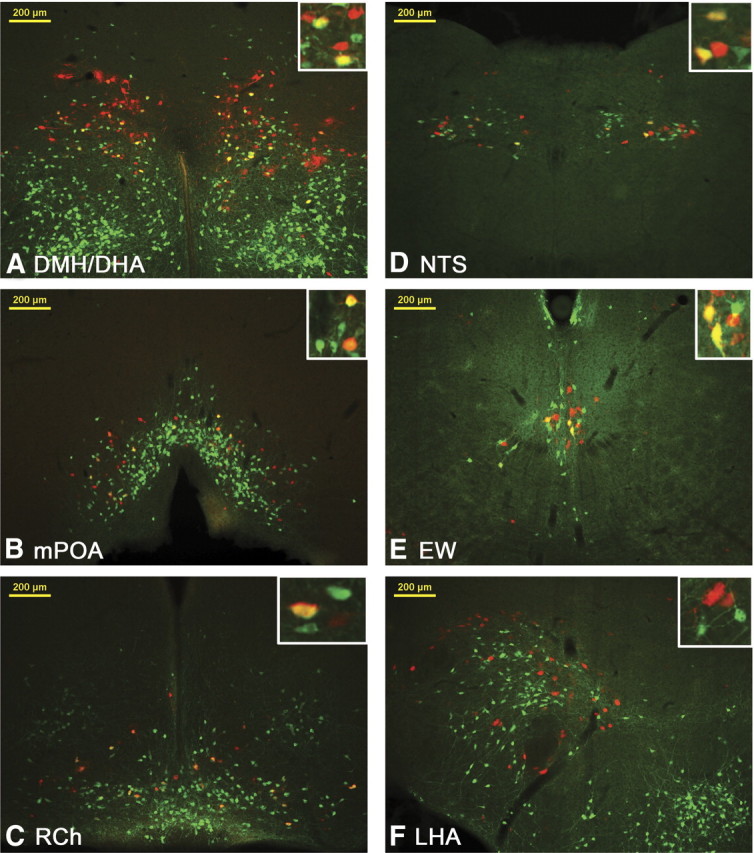
Representative images from LepRbEGFP mice 96 h after PRV infection of the BAT. Transsynaptically and retrogradely PRV (red stain) traced LepRbEGFP (greed stain) neurons are found in the DMH/DHA (A), mPOA (B), RCh (C), NTS (D), and EW (E). Other PRV- and LepRb-positive sites, such as the lateral hypothalamic area (LHA, F), did not show colocalized neurons.