
Figure 8.
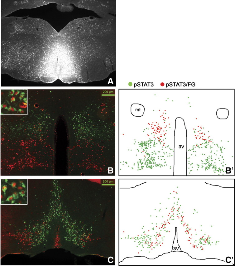
Stereotaxic injection of the retrograde tracer FG into the RMR (A, n = 4) and immunohistochemical detection of retrogradely labeled FG (green) in LepRb neurons (leptin-induced pSTAT3, red) in the DMH/DHA (B) and mPOA (C). Schematic drawings of double-labeled FG/pSTAT3 neurons (red dots) in contrast to non-FG-labeled pSTAT3 neurons (green dots) in the DMH/DHA (B′) and mPOA (C′). 3V, Third ventricle.