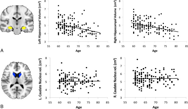
Figure 3.
A, Example of hippocampal segmentation and scatter plots showing decline in volume of the left and right hippocampus between 59 and 81 years of age (significant at p < 0.001). B, Example of caudate nucleus segmentation and scatter plots showing no significant decline in volume of the caudate nucleus between 59 and 81 years of age.

