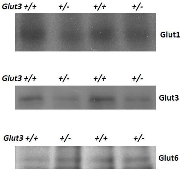
Figure 1. Expression of Glut1, Glut3, and Glut6 in brain homogenate from Glut3+/− mice.
Shown here are representative immunoblots of brain homogenates subjected to PAGE and probing of resulting membranes with antibodies against mGlut3, hGLUT1, and hGLUT6. Digital image analysis quantified the mean expression of Glut3 at 52% of the Glut3+/+ littermates. The expressions of Glut1 and Glut6 in Glut3+/− brain were quantified at 100% and 100%, respectively.

