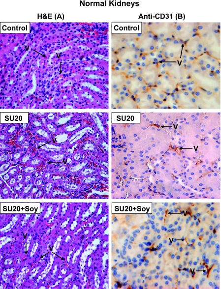
Figure 4.
Histology of normal kidneys from mice treated with sunitinib and soy. The normal contralateral kidneys from control mice or mice treated with SU20 and SU20 combined with soy (SU20 + Soy), obtained on day 27 from experiments described in Figure 2, were processed for histology and H&E staining (A) and anti-CD31 immunostaining (B). Only alterations induced by SU20 or soy can be observed in these left kidneys because they were not in the field of radiation. The normal kidney from control mice showed intact, regular, and thin blood vessels (A, B). The vessels in normal kidneys of mice treated with SU20 showed mild dilatation (A, B). In contrast, the vessels of normal left kidneys treated with systemic SU20 and soy looked thinner and more regular (A) as confirmed by anti-CD31 staining of endothelial cells (B).

