Figure 4.

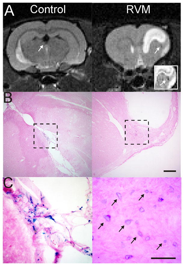
Iron-oxide labeled NSCs are still visible on MRI at 58 weeks. A) A control animal implanted with NSCs within the ventricle still exhibits MRI hypointensities within the ventricle (arrow) at 58 weeks after implantation. An RVM animal that had an intra-striatal implantation of NSCs clearly shows an MR visible hypointensity adjacent to the lesion (arrow). Inset clearly identifies a loss of T2 signal (arrow) adjacent to the lesion site. B) A histological section from the control animal shows no lesion but iron labeled mNSCs within the ventricle. In contrast, the RVM animal has iron labeled mNSCs adjacent to the lesion. Cal bar = 0.5mm. C) A high power photomicrograph (40X) allows clear visualization of iron labeled cells (arrows) still within the choroid plexus of the ventricle even at 58 weeks, whereas the mNSCs are neighboring the lesion in the RVM animals Cal bar = 100um.
