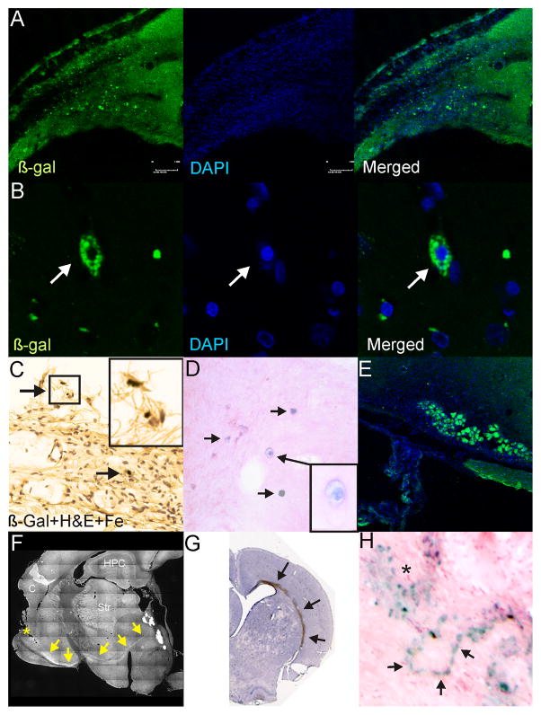
Figure 5.
Immunohistochemical and histological assessment of NSCs at 58 weeks after implantation. A) Dual labeled fluorescent staining revealed that numerous β-galactosidase (β-gal) positive cells could be observed in the vicinity of the ischemic lesion. B) High power (100X) examination of these cells revealed that β-gal was localized within the cytoplasm and these cells were healthy with DAPI staining in the nucleus. C) Similarly, at more ventral locations, similar iron and β-gal stained cells could be observed (arrows). Inset: high power magnification illustrates iron containing cells. D) Prussian blue staining followed by H&E counterstaining also demonstrates numerous iron containing cells within the parenchyma adjacent to the injured tissues. E) In many sites adjacent to the lesion large groups of β-gal stained cells could be observed, suggesting the possibility that many of these cells may replicate in the vicinity of the injury. F) BDA injection (*) in a subset of animals revealed unusual anterograde transport within the brains of these animals. The appearance of a robust stained pathway at the base of the striatum was noted. G) Gross histological specimen showing robust staining within the corpus callosum of an animal following parenchymal injection at 58 weeks after implantation. Robust β-gal positive staining could be observed on the side of the HII injury. H) Similarly, even with Prussian blue staining numerous clusters (*) of iron labeled NSCs could be observed, but chain-like series of iron labeled cells could also be seen.

