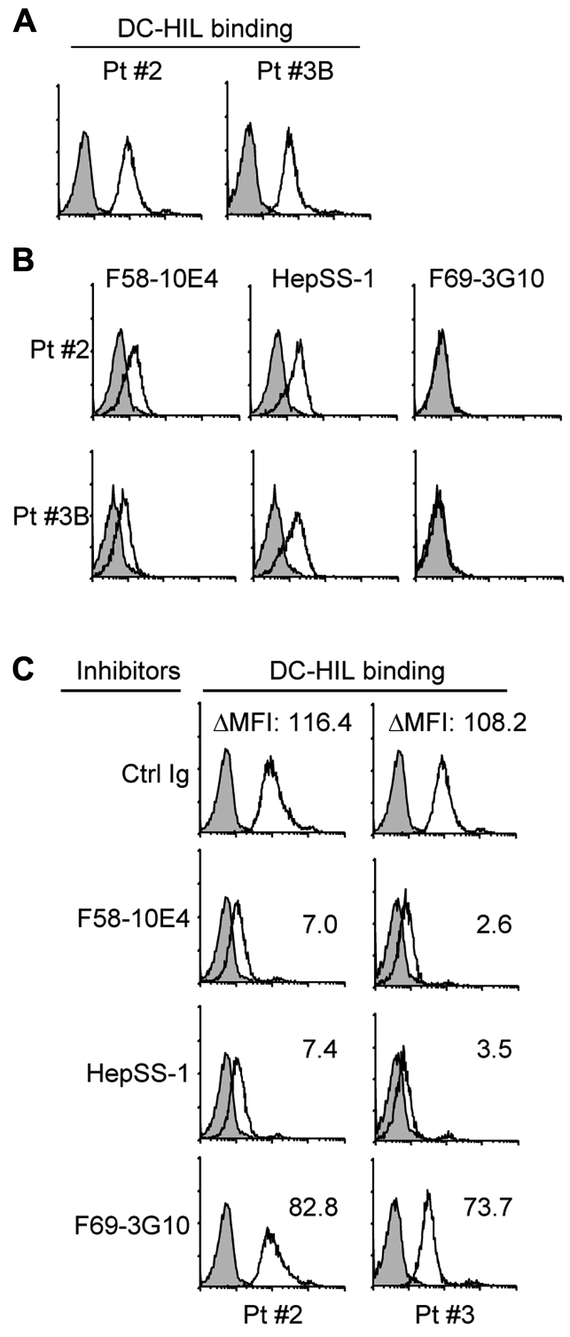
Figure 3.

PBMCs of patients with SS use distinct HS moieties to bind DC-HIL at high levels. PBMCs freshly isolated from patients with SS (Pt. 2 with Vβ+ cells at 93%, in which SD-4+ cells were 95%, and Pt. 3B with Vβ+ cells at 98.3%, in which SD-4+ cells were 96%) were assayed by flow cytometry for DC-HIL binding (A) and expression of HS epitopes (B) as in Figure 2. (C) DC-HIL binding by these cells was blocked by pretreatment with 40 μg/mL anti–HS mAb or control Ig (IgM plus IgG). DC-HIL binding is assessed by MFI left after subtracting MFI of control staining from MFI of positive staining (ΔMFI).
