Figure 4.
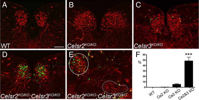
Celsr2 and Celsr3 are required for FBM neuron survival. A–F, FBM neurons shown with anti-Isl1 (red) were stained for activated (cleaved) caspase-3 (green) (A–E) at E12.5. No apoptotic cells were found in WT (A) and Celsr2KO/KO mice (B). In Celsr3KO/KO (C), a few FBM neurons were positive for activated caspase-3. In Celsr2KO/KO;Celsr3KO/KO (D–F), the number of caspase-3 positive cells increased dramatically in FBM neurons located in the medial region (circle), but not when they finish lateral migration (dotted contour). F, Cell death, estimated as the ratio of green to red signals (ImageJ software), was 5% in Celsr3KO/KO and 50% in Celsr2KO/KO;Celsr3KO/KO samples. ***p < 0.001, Student's t test, Bonferroni correction. Error bars are SD. Scale bar (in A) A–E, 100 μm.

