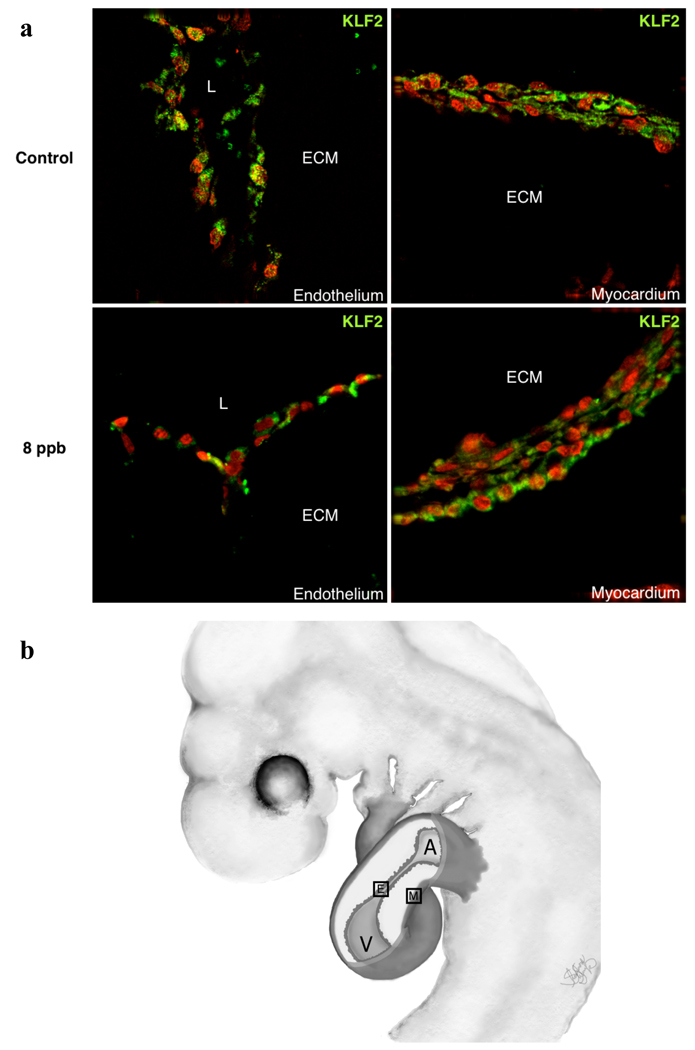
Figure 3.
KLF2 Antibody Expression in HH17 Whole Hearts collected after approximately 24hr exposure of 8 ppb TCE. KFL2 Antibody expression can be observed in both the endothelium and myocardium of the developing chick heart in both the control and 8 ppb TCE treated samples. Staining in TCE-treated endothelia appears to show an increase in extra-nyclear distribution of the protein.

