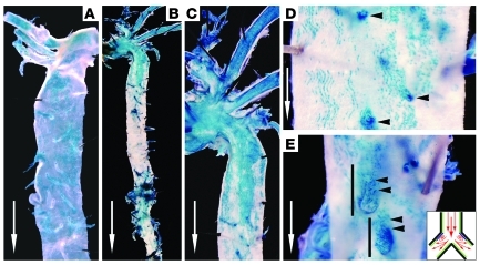
Figure 1. Tie1 expression at shear stress–specific sites in the adult aorta.
Representative en face X-gal–stained, whole-mount distal aorta of 4-week-old (A) and 12-week-old (B–E) Tie1-LacZ mice. (A) Pervasive endothelial Tie1-LacZ expression in the aortic arch, branch arteries, and descending aorta wall of immature 4-week-old mouse. (B) Low-magnification view of aorta (aortic arch and thoracic and abdominal aorta) and (C) higher-magnification view depicting endothelial Tie1-LacZ expression in the aortic arch diminishing in the thoracic aorta as laminar flow is restored. (D) High-magnification image of thoracic aorta wall, illustrating endothelial Tie1-LacZ expression at ostia of vertebral artery branches (arrowheads). (E) High-magnification image of abdominal aorta wall, showing distinct X-gal staining at the outer walls of renal artery bifurcations (arrowheads). Inset: illustration of proatherogenic, disturbed flow as experienced at aortic branch points. White arrows indicate direction of blood flow. Original magnification, ×10 (B); ×20 (A and C); ×40 (D and E).

