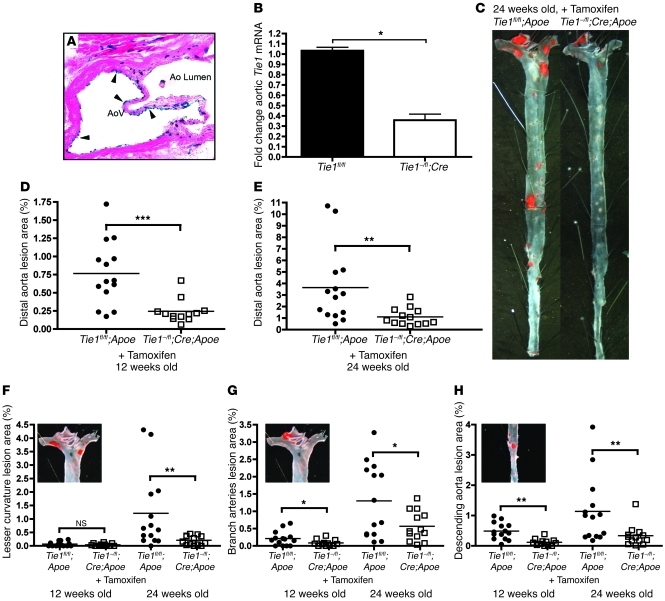
Figure 4. Endothelial-specific Tie1 deletion reduces atherosclerosis burden.
(A) Representative H&E-stained aortic valve showing endothelial-specific LacZ expression (arrowheads) from tamoxifen-treated SCL-ERT-Cre;Rosa26R-LacZ mouse. Original magnification, ×100. (B) RT-PCR analysis of aortic Tie1 levels from tamoxifen-treated Tie1–/fl;SCL-ERT-Cre mice (65% reduction, P < 0.05). (C) Representative Sudan IV–stained distal aorta from Tie1–/fl;SCL-ERT-Cre;Apoe–/– and Tie1fl/fl;Apoe–/– mice demonstrated lesion reductions in Tie1-deleted mice. (D and E) Mean aortic lesion area in tamoxifen-treated Tie1–/fl;SCL-ERT-Cre;Apoe–/– and Tie1fl/fl;Apoe–/– mice analyzed at (D) 12 weeks (68% reduction, P < 0.0005) and (E) 24 weeks (70% reduction, P < 0.006). (F–H) Atherosclerotic lesion areas in Tie1-deleted mice, as assessed in 3 regions of the aorta, showed reduction of atherosclerosis in all regions of disturbed flow. Insets show respective regions of lesion analysis. Shown are mean atherosclerotic lesion areas at (F) lesser curvature of the aortic arch (12 weeks, 0.066% ± 0.023% vs. 0.038% ± 0.014%, P = NS; 24 weeks, 1.20% ± 0.379% vs. 0.206% ± 0.048%, 82% reduction, P < 0.01); (G) aortic arch branch arteries, including brachiocephalic, left common carotid, and left subclavian arteries (12 weeks, 0.211% ± 0.055% vs. 0.086% ± 0.026%, 56% reduction, P < 0.05; 24 weeks, 1.29% ± 0.283% vs. 0.568% ± 0.119%, 59% reduction, P < 0.05); and (H) descending aorta (12 weeks, 0.487% ± 0.074% vs. 0.118% ± 0.032%, 70% reduction, P < 0.01; 24 weeks, 1.13% ± 0.291% vs. 0.333% ± 0.097%, 75% reduction, P < 0.01). Data points denote individual animals, and horizontal bars indicate group average. *P < 0.05; **P < 0.01; ***P < 0.001.

