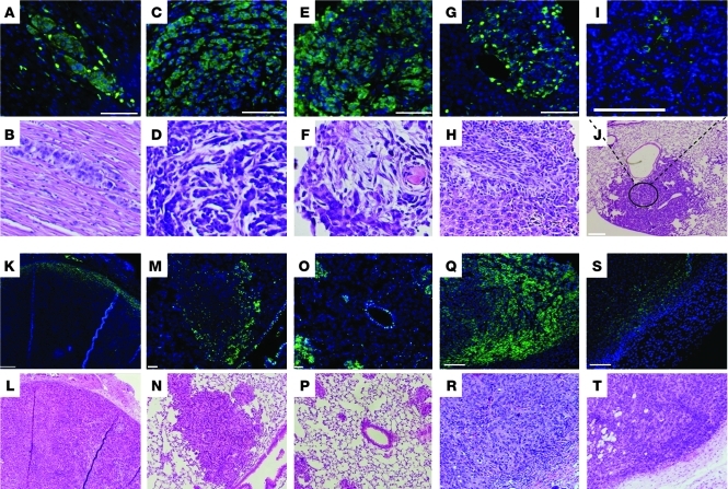
Figure 1. CD133 expression in primary and metastatic tumors in KrasLA1/+p53R172HΔG/+ mice and in syngeneic mice injected with 344SQ cells.
CD133 staining (green) in metastases to (A and B) heart, (C and D) diaphragm, (E and F) chest wall, and (G and H) liver and (I and J) a primary lung tumor from KrasLA1/+p53R172HΔG/+ mice, (K and L) in a primary subcutaneous tumor and (M and N) large and (O and P) small lung metastases from syngeneic mice injected with 344SQ cells, and in a primary subcutaneous tumor in mice injected with (Q and R) CD133hi and (S and T) CD133lo 344SQ tumor cells. Sections were costained with DAPI (blue). Under each CD133/DAPI-stained panel is a panel with hematoxylin and eosin staining of an adjacent tissue section. Original magnification, ×20 (A–I and M–T); ×4 (J–L). Scale bars: 100 μm (A–I, L, N, and P–T); 200 μm (J and K); 50 μm (M and O).

