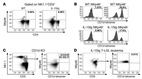
Figure 3. NKp46+ NKT cells are CD4– non-CD1d-restricted NKT-like cells.
(A) Splenic cells from WT and IL-15tg mice were stained with NK1.1, CD3, CD4, and NKp46 mAbs. Cells were first gated on NK1.1+CD3+, followed by an assessment of NKp46 and CD4 expression. Percentages of cells are only shown for the top right quadrants. (B) Splenic cells from WT and IL-15tg mice were stained with the mAbs of NK1.1, CD3, NKp46, CD19, and CD1d tetramer loaded with the α-C-galactosylceramine analog PBS-57. NK1.1+CD3+CD19– cells were first gated and then examined for NKp46 and CD1d expression. Percentages of cells positive for CD1d tetramer binding are shown. (C) Splenic cells from CD1d-deficient (CD1d KO) mice were stained with NK1.1, CD3, CD19, NKp46, and CD1d tetramer mAbs. CD19+ cells (data not shown) were first gated out, and the remaining cells were examined for the NK1.1+CD3+ population, followed by gating and an analysis of NKp46 and CD1d expression. Percentages of cells staining positive for all 4 quadrants are shown. (D) Splenic cells from IL-15tg T-LGL leukemia mice were stained with CD3, NKp46, CD19, and CD1d tetramer mAbs. CD19+ cells (data not shown) were first gated out, and the remaining cells were examined for the NKp46+CD3+ population, which was then assessed for CD1d expression. Data are representative of 1 out of at least 3 mice showing similar results. The percentage of cells staining positive in top right quadrant is shown for the right panel.

