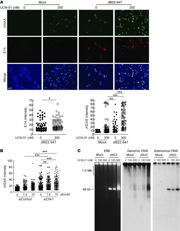
Figure 5. Loss of Chk1 activity increases genomic DNA damage.
(A) A2780CP cells were infected with dl922-947 (MOI 7.5) and treated with UCN-01 6 hours later. 48 hpi, cells were fixed, stained for expression of E1A and γH2AX, counterstained with DAPI, and imaged. Fluorescence intensity was assessed in more than 100 cells per condition using ImageJ software. Each point represents a single cell, and each red bar represents the median. Original magnification, ×40. *P < 0.05, ***P < 0.0001; Mann-Whitney test. (B) IGROV1 cells were transfected with Chk1 (siChk1) or scrambled control siRNA (siControl) oligonucleotides 24 hours prior to infection with dl922-947 (MOI 7.5 and 15). Cells were fixed 24 hours thereafter, stained for expression of γH2AX, and counterstained with DAPI. Fluorescence intensity was assessed as before. **P < 0.01, ***P < 0.0001; Mann-Whitney test. (C) A2780CP cells were infected with dl922-947 (MOI 7.5) for 48 hours, with or without treatment with UCN-01. DNA was extracted and subjected to neutral PFGE and probed with HRP-labeled genomic DNA or adenovirus type 5 probe.

