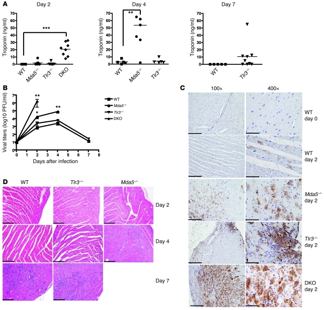
Figure 2. MDA5 controls EMCV-D infection in the heart.
WT and KO mice were infected with 103 PFU of EMCV-D. Serum and heart (n ≥ 6 per time point, 3 independent experiments) were harvested at days 2, 4, and 7 from surviving mice and were evaluated for (A) troponin levels by ELISA and (B) virus titer by plaque assay or fixed in formalin and paraffin embedded for histology. Tissue sections were stained for EMCVpol antigen by immunohistochemistry (C) (left column: original magnification, ×100 magnification; scale bars: 200 microns; right column: original magnification, ×400; scale bars: 50 micron) or stained by H&E and evaluated for pathology (D) (original magnification, ×100; scale bar: 200 micron). In C, strong and diffuse EMCVpol reactivity is particularly evident in the myocardium of Mda5–/– and DKO; in contrast, heart from WT and Tlr3–/– animals showed only mild (WT) or focal (Tlr3–/–) staining, which is shown in detail in the right panel. In D, areas of myocarditis are observed in WT and Tlr3–/– on day 7 PI as well as in Mda5–/– day 4 PI; only mild inflammation is observed in Tlr3–/– day 4 PI. Statistical significance was calculated by 2-tailed Student’s t test and is indicated as follows: *P < 0.05; **P < 0.01; ***P < 0.001.

