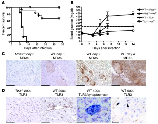
Figure 4. Stromal MDA5 and hematopoietic cell TLR3 protect against EMCV-D infection.
(A and B) BM chimeras were generated between WT and Mda5–/– (n = 10 each) and WT and Tlr3–/– animals (n = 12 each) in 2 independent experiments. Chimeras were infected with 103 PFU of EMCV-D and evaluated for (A) survival and (B) blood glucose. Mda5–/–→WT and WT→Tlr3–/– chimeras survived infection, WT→Mda5–/– chimeras succumbed on day 6, and Tlr3–/–→WT chimeras succumbed on day 14 with 67% surviving infection. (C) MDA5 expression in the pancreas (original magnification, ×200; scale bars: 100 micron). Fixed tissue sections were made from Mda5–/– and WT pancreas on days 0, 2, and 4 after EMCV infection and stained with anti-MDA5 (n = 3). MDA5 is expressed in islets before infection and induced in both islets and exocrine pancreas after infection. (D) TLR3 expression in the pancreas. Frozen tissue sections were made from WT and Tlr3–/– pancreas from uninfected animals (first, second, and fourth panels) or from WT pancreas 12 hours after EMCV infection (third panel). Sections were stained with anti-TLR3 (brown) or costained with anti-TLR3 (brown) and synaptophysin (blue) to visualize expression in the islets (third panel) and are shown at different magnifications as indicated (original magnification, ×200, scale bars: 100 micron; original magnification, ×400, scale bars: 50 micron; original magnification, ×600, scale bars: 33 micron) (n = 3). TLR3 expression is found in the islets as well as in duct epithelial cells, vascular endothelial cells and interstitial stromal cells (arrowheads indicate TLR3+ interstitial cells).

