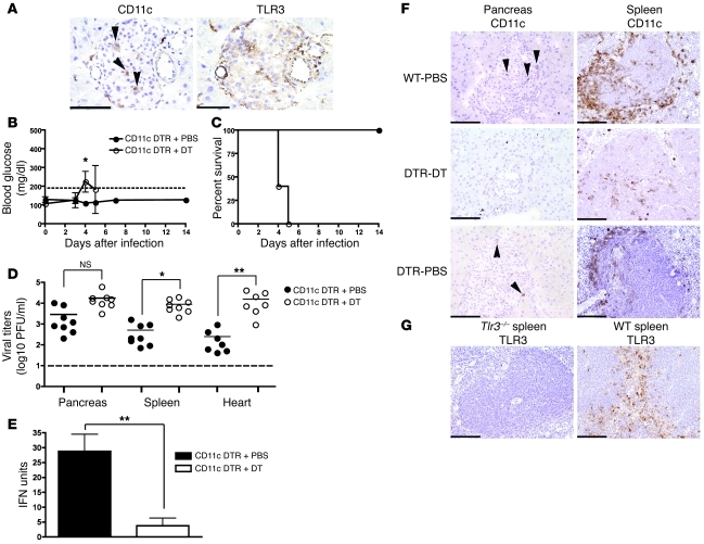
Figure 5. CD11c+ DC control EMCV-D infection and development of diabetes.
(A) Identification of CD11c+ DCs in the islets. Serial frozen sections were made from the pancreas of uninfected WT animals and stained with anti-CD11c (left panel) and anti-TLR3 (right panel) (original magnification, ×400; scale bar 50 micron) (n = 3). Arrowheads indicate CD11c+ cells that are also TLR3+. (B–E) CD11c+ DC are required for protection from EMCV-D–induced diabetes. CD11c-DTR mice were treated with PBS (n = 8) or DT (n = 8), then monitored for (B) blood glucose, (C) survival, (D) organ viral titers in the pancreas, heart, and spleen, and (E) serum IFN-I after EMCV-D infection. (F) Frozen sections were prepared from WT and CD11c-DTR PBS and DT-treated mice 48 hours after treatment. Immunohistochemical analysis of CD11c+ cells in the pancreas and spleens of these animals revealed depletion primarily in the spleen after DT treatment (original magnification, ×200; scale bars: 100 micron) (n = 3). Arrowheads indicate CD11c+ cells in the pancreas. (G) Frozen sections of spleens from WT and Tlr3–/– mice were used to perform immunohistochemical evaluation of TLR3 expression in the marginal and periarteriolar zones of the spleen (original magnification, ×200, scale bars: 100 micron) (n = 3). Statistical significance was calculated by 2-tailed Student’s t test and is indicated as follows: *P < 0.05; **P < 0.01.

