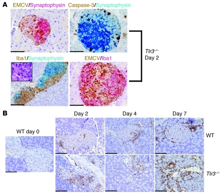
Figure 7. EMCV-D–induced diabetes in Tlr3–/– mice is characterized by islet infection, apoptosis, and myeloid cell infiltrate.
Pancreas tissue samples from WT or Tlr3–/– mice were harvested at days 0, 2, 4, or 7 PI as indicated, fixed in formalin, and paraffin embedded. (A) Sections were costained with anti-EMCVpol (brown) and anti-synaptophysin (red) (top left panel); active caspase-3 (brown) and anti-synaptophysin (blue) (top right panel); anti–Iba-1 (brown) and anti-synaptophysin (blue) with H&E-stained insert (bottom left panel); or anti-EMCVpol (brown) and anti–Iba-1 (red) (bottom right panel) (left panels: original magnification, ×200, scale bars: 100 micron, right panels: original magnification, ×400, scale bars: 50 micron). By morphology, Iba-1+ cells correspond to large cells with irregular nuclei resembling macrophages (A, bottom left panel insert). As demonstrated by double immunohistochemistry, these islets contain numerous apoptotic cells and Iba1+ cells that react with antibodies against EMCVpol. (B) To visualize myeloid cell infiltrates, sections from WT and Tlr3–/– pancreas were stained with anti–Iba-1 (original magnification, ×200; scale bars: 100 micron). In WT pancreas, Iba-1+ cells are found at the periphery of the islets, whereas In Tlr3–/–, Iba-1+ cells infiltrate the islet.

