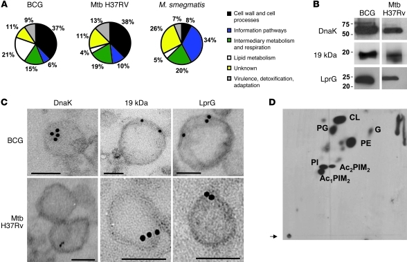
Figure 3. Mycobacterium vesicle–associated proteins and lipids.
(A) Pie charts showing the BCG, Mtb, and M. smegmatis vesicle–associated proteins grouped into functional categories as in TubercuList, BoviList, and SmegmaList. Note that the “lipid metabolism” functional category is increased in BCG vesicles and that “cell wall and cell processes”–associated proteins are overrepresented in both BCG and Mtb compared with M. smegmatis. (B) Vesicles from the supernatant of the BCG culture were loaded onto a SDS-polyacrylamide gel, blotted on a nitrocellulose membrane, and incubated with mAbs with the indicated specificities. (C) ImmunoGold electron microscopy of thin sections of BCG and Mtb H37Rv vesicles treated with the mAbs specific for the indicated proteins and detected with a 10-nm IgG gold-labeled anti-mouse antibody. Scale bars: 100 nm. (D) Total lipids of vesicles isolated from 14C-acetate–labeled cells were extracted with chloroform/methanol (2:1, v/v). Lipid extracts were separated by 2D TLC by applying an amount of lipids corresponding to 10,000 DPM per TLC plate and by using the solvent system E for separation of polar lipids. PG, phosphatidylglycerol; G, glycolipid; Ac1PIM2, monoacyl phosphatidylinositol dimannoside; Ac2PIM2, diacyl phosphatidylinositol dimannoside. Arrow indicates the origin.

