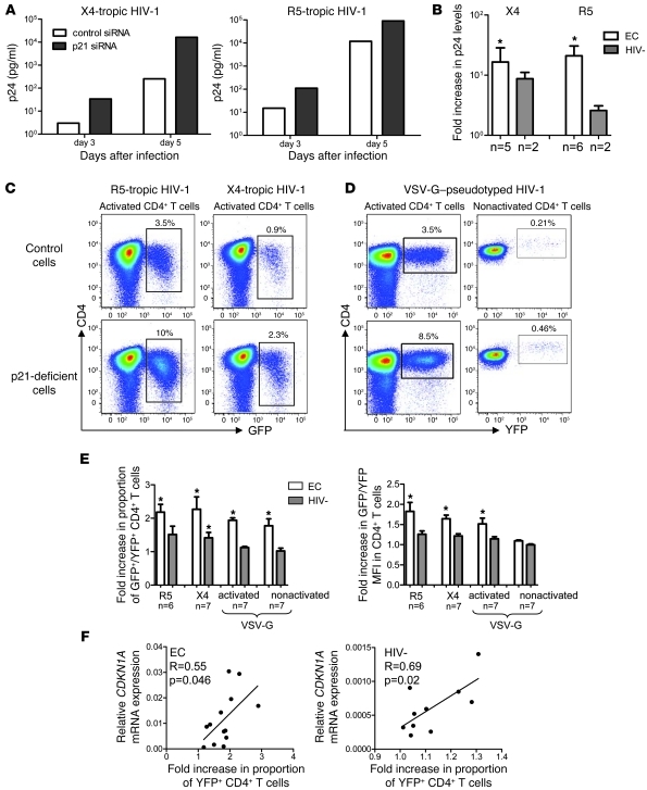
Figure 4. Inhibition of p21 enhances HIV-1 replication in CD4+ T cells.
(A and B) Activated CD4+ T cells from elite controllers or HIV-1–negative persons were infected with X4- or R5-tropic primary HIV-1 isolates in the presence of p21-specific or control siRNA; HIV-1 replication was assessed by p24 antigen levels in culture supernatants. (A) Representative example from an HIV-1 elite controller. (B) Fold increase (mean and SD) of p24 levels in p21-deficient versus control cells in indicated persons. (C–F) Flow cytometric assessment of HIV-1 replication in CD4+ T cells after p21 inhibition. CD4+ T cells were infected with GFP-encoding X4- or R5-tropic HIV-1 strains in the presence of p21-siRNA or control siRNA or with a YFP-encoding VSV-G–pseudotyped HIV-1 vector in the presence of a pharmacological p21 inhibitor or the carrier DMSO. (C and D) Representative dot plots from an elite controller. Percentages indicate the proportion of gated CD4+ T cells. (E) Fold increase (mean and SD) in the proportion of GFP+/YFP+ cells or in GFP/YFP mean fluorescence intensity in p21-deficient cells compared with controls. White bar, EC; Grey bar, HIV-1. (F) Correlation between CDKN1A mRNA expression in HLA-DR– CD4+ T cells from elite controllers and HIV-1–negative persons and corresponding fold increases of YFP+ cells after p21 inhibition. *P < 0.05; p21-deficient cells versus control cells treated with unspecific siRNA or SMSO; paired Wilcoxon test. Statistical comparison was performed using Student’s t test.

