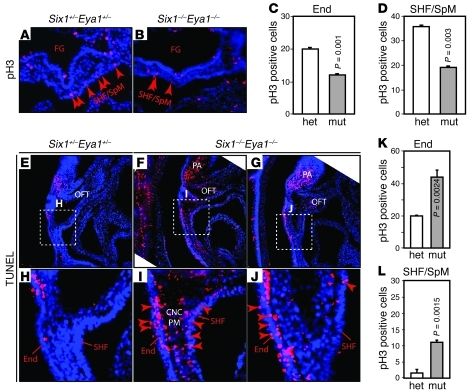
Figure 3. Six1 and Eya1 are required for cell proliferation and survival in PA and OFT.
(A–D) Anti-pH3 immunostaining (red) of E9.5 transverse sections to label proliferating cells. Quantification of results are shown in C and D. FG, foregut; mut, Six1–/–Eya1–/– mutant; het, heterozygous control. n = 3. (E–L) TUNEL staining of sagittal sections revealed increased cell death in pharyngeal endoderm, SHF/SpM, and pharyngeal mesenchyme (PM) that contained CNCs and mesoderm. Boxed regions in E–G are enlarged in H–J. Original magnification, ×200 (A, B, and E–J). n = 3. P values were determined by Student’s t test.

