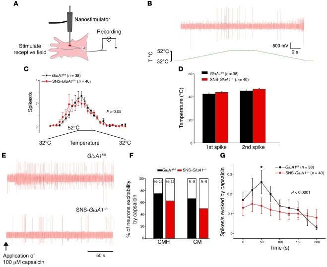
Figure 3. Electrophysiological analysis of peripheral nociceptive fiber responses to nociceptive stimuli in GluA1fl/fl mice and SNS-GluA1–/– mice.
(A) Scheme of the skin-saphenous nerve preparation and the recording setup (see Methods for details). (B and C) Analysis of responses of C-mechanoheat nociceptors to heat ramps. (B) A typical example of spike activity in response to a heat ramp. T, temperature. (C) Average spike activity from a population of nociceptors. (D) The threshold temperature for evoking first and second spikes. (E–G) Analysis of responses of C-mechanoheat (C-MH) and C-mechano (CM) types of nociceptors to application of capsaicin (100 μM) to the receptive field in the skin. (E) Typical examples of capsaicin-evoked spikes in SNS-GluA1–/– and GluA1fl/fl mice. (F) Cumulative summary of the incidence of capsaicin-sensitive nociceptors. (G) Summary of the magnitude of capsaicin-evoked action potentials in nociceptors of SNS-GluA1–/– and GluA1fl/fl mice. No significant differences were seen between genotypes in C, D, and F. Two-way ANOVA for repeated measures revealed significant differences in the cumulative curves representing SNS-GluA1–/– and GluA1fl/fl mice (P < 0.001). *P < 0.05 for the time point indicated (post-hoc Bonferroni test).

