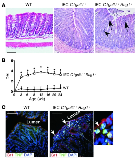
Figure 2. Adaptive immunity is not required for the initiation of colitis in IEC C1galt1–/– mice.
(A) Representative H&E-stained colonic sections indicate similar colonic inflammation in IEC C1galt1–/– mice with and without Rag1 deficiency at 2 weeks. (B) Clinical DAI (mean ± SD, n = 10 mice/group) of IEC C1galt1–/– mice based on diarrhea, fecal occult blood, and rectal prolapse. *P < 0.05. (C) Cryosections of IEC C1galt1–/–Rag1–/– colon stained with mAbs to myeloid cells (Gr1) and TNF at 3 weeks. Arrows mark infiltrates in epithelium. Inset (original magnification, ×400) highlights TNF-positive myeloid cells. Scale bars: 100 μm in A, 50 μm in C. Data are representative of at least 3 experiments.

