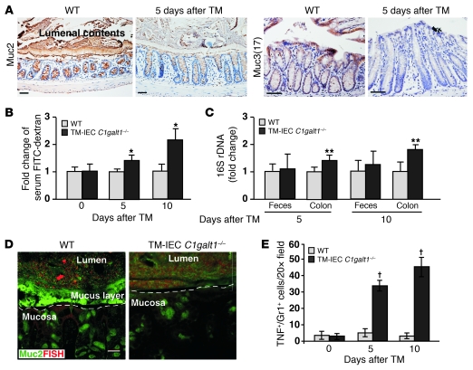
Figure 6. Absence of core 1–derived O-glycans impairs the levels of intestinal mucins and the integrity of intestinal barrier function.
(A) Representative images of immunohistochemical staining (brown) with O-glycosylation–independent antibodies to different mucins in colon sections of WT and TM-IEC C1galt1–/– mice 5 days after TM. Scale bars: 100 μm. (B) Serum concentrations of FITC-dextran were measured 4 hours after oral administration to WT and TM-IEC C1galt1–/– mice at different time points after TM induction (mean ± SD, n = 6). *P < 0.008. (C) Relative amount of bacterial 16S rDNA detected by real-time PCR using two independent sets of 16S universal primers. Data are expressed as the fold difference between WT and TM-IEC C1galt1–/– mice (mean ± SD, n = 6). The average 16S rDNA value of WT mice was expressed as 1. **P < 0.02. (D) Colon sections of 7-week-old mice 10 days after TM induction were probed with an Alexa Fluor 555–conjugated general bacterial 16S rDNA probe for FISH (red) and costained with a FITC-labeled glycosylation-independent polyclonal antibody to Muc2 (green). Dashed lines mark epithelial surfaces. Scale bar: 20 μm. (E) Quantification of immunofluorescence images of 7-week-old WT and TM-IEC C1galt1–/– colon sections stained with mAbs to myeloid cells (Gr1) and TNF (mean ± SD, n = 5). †P < 0.001.

