
Figure 3.
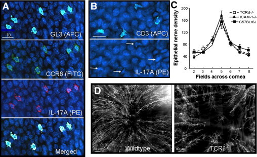
Corneal nerve regeneration in TCRδ−/− and ICAM-1−/− mice. A: GL3+ cells (pseudocolor light blue) in the paralimbal epithelium of wild-type mice at 18 hours after central epithelial abrasion. These cells were also positive for CCR6 (green) and IL-17A (red). Blue nuclei of basal epithelial cells stained with DAPI. B: Cornea from a TCRδ−/− mouse at 18 hours after central epithelial abrasion showing CD3+ cells (pseudocolor light blue) in the paralimbus. The cornea was also stained with anti-IL-17A-PE, but in contrast to wild-type corneas, the CD3+ cells (arrows in lower panel) were not labeled. C: Epithelial nerve fiber density in seven microscopic fields across the cornea from paralimbus to paralimbus comparing three strains of unwounded, age-matched male mice. No statistical differences were found among the strains. D: Photomontages of the central cornea of a wild-type and an age- and sex-matched TCRδ−/− mouse 96 hours after central epithelial abrasion. Nerves were stained with anti-tubulin III-PE.