
Figure 4.
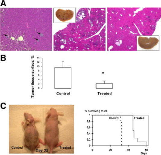
In vivo effects of rapamycin on intrahepatic cell growth. Rapamycin (1.5 mg/kg daily) was administrated from day 8, to allow engraftment of tumor cells in the liver, to day 25. A, left: histologic examination of day 8 control mice: small early nodules are visible in the liver (arrows); middle and right: histologic examination of control and treated mice sacrificed at day 25: many nodules of variable size are present in the liver of control mice (middle), whereas only few small nodules are visible in rapamycin-treated mice (right) (H&E staining, original magnification, ×4); inserts: macroscopic view showing a strong decrease of the number of visible liver nodules. B: Morphometric analysis: the total tumor tissue surface is strongly reduced on rapamycin treatment (*P < 0.05). C: Survival study: Mice received either vehicle or rapamycin injection (1.5 mg/kg daily) from day 8 after STC-1 intrasplenic injection. Left: morphologic aspect of a control and a rapamycin-treated mouse 32 days after STC-1 intrasplenic injection. Right: Kaplan-Meier curve showing a significant difference of survival between control and rapamycin-treated mice (P < 0.002).