
Figure 2.
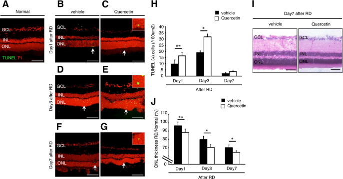
Quercetin-induced photoreceptor apoptosis 3 days after retinal detachment (RD). A–G: Transferase-mediated dUTP nick-end labeling (TUNEL) assay in the normal (A), vehicle (B, D, and F) and quercetin (C, E, and G) at 1, 3, and 7 days after RD. TUNEL (+) cells (arrows) were observed in the outer nuclear layer (ONL) (C, E, and G). Insets are higher magnification of arrows at panel C, E, and G). H: Quantification of TUNEL (+) cells by immunohistochemistry. Note that the number of TUNEL (+) cells was significantly lower in the vehicle than in the quercetin (*P < 0.01; **P < 0.05). I: Representative photomicrographs of RD at 7 days. J: Relative ratio of the ONL thickness in each group. In the both groups, ONL thickness was decreased with time. However, ONL thickness in the quercetin group was significantly decreased compared with the vehicle group of them (*P < 0.01, **P < 0.05). Each column represents the mean ± SD of four independent experiments. Scale bars (A–G, I) = 100 um. GCL, retinal ganglion cell layer; INL, inner nuclear layer.