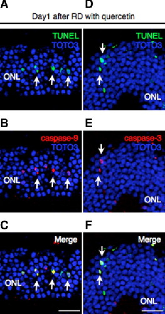
Supplementary Figure S2.

(A–F) In the quercetin group, intensive staining of cleaved caspase-9 (B) (red) and caspase-3 (E) (red) was detected with laser-capture microdissection (A and D) (green) assay in the photoreceptor cells (arrows). Bars, 50 um. ONL, outer nuclear layer; RD, retinal detachment.
