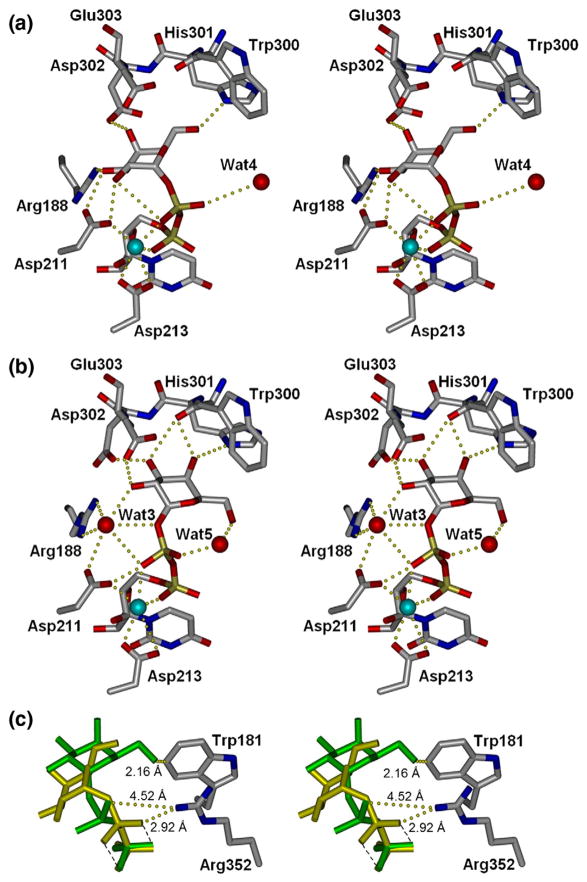
Fig. 4.
Stereoviews of enzyme bound to UDP-galactose. (a) Wild-type GTB9 and (b) Cys-to-Ser mutant enzymes. Cyan spheres represent Mn2+. (c) Compared to wild type (yellow), this novel UDP-Gal conformation (green) displaces the closed conformation of the internal loop by steric hindrance of Trp181 and inhibition of C-termini closure by rotation of β-phosphate out of Arg352 access.

