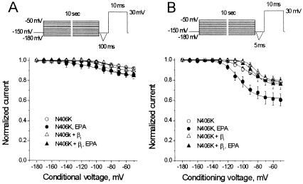
Figure 7.
Voltage-dependent suppression of INa by EPA in HEK293t cells coexpressing N406K and the β1 subunit of hH1α Na+ channels. (A) Whole-cell currents were normalized by INa recorded with the conditioning voltage of −180 mV for their corresponding controls. The experimental protocol is shown in the Inset. Currents were elicited by 10-ms test pulses to 30 mV following a 10-s conditioning pulse varying from −180 mV to −50 mV with 10-mV increments. A 100-ms interval was inserted between the conditioning pulse and the test pulse. The membrane potential was held at −150 mV, and the pulse rate was 0.1 Hz. EPA at 5 μM did not show a significantly voltage-dependent suppression of INa for N406K alone or N406K plus β1 with the protocol of a 100-ms recovery interval. (B) Voltage-dependent suppression of INa in the presence of 5 μM EPA is shown. The Inset is the voltage protocol with a recovery interval of 5 ms. With this protocol, 5 μM EPA did not produce a significant voltage-dependent suppression of INa in HEK293t cells coexpressing N406K and β1. The data were fit with a Boltzmann equation. Each data point represents the average value of at least eight individual cells.

