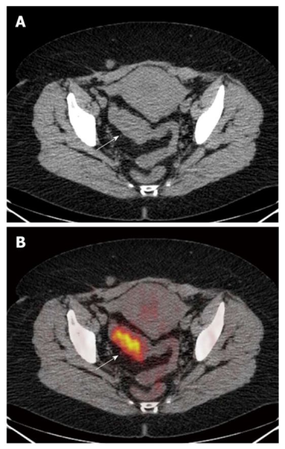
Figure 1.

The computed tomography (A) and fused positron emission tomography/computed tomography (B) images. A: Neoplastic thickening of sigmoid colon walls, which show regular profiles with normal appearance of perilesional fat (arrow); B: Uptake of 18-FDG (arrow) (SUVmax 11) in the absence of extra-parietal radio-tracer uptake. The lesion was correctly classified as ≤ T2.
