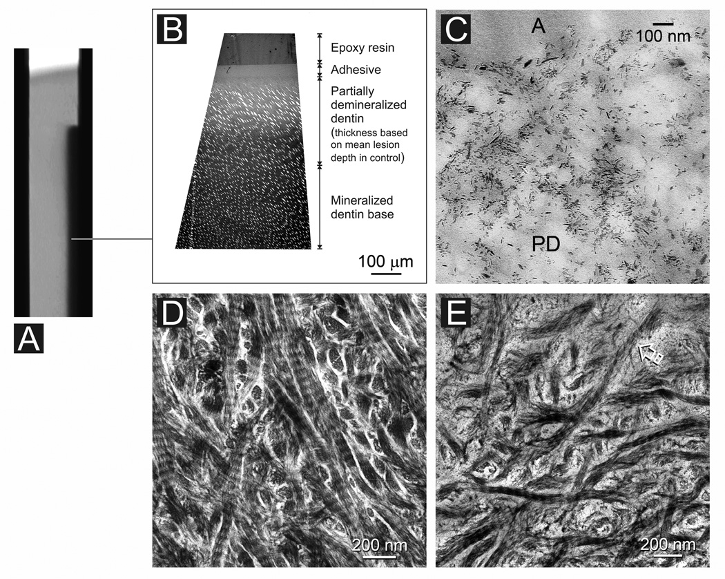
Fig. 3.
Characterization of an adhesive infiltrated artificial caries-like dentin lesion after 4 months of immersion in simulated body fluid (control). A. A stacked 2-D micro-CT image representing the lesion profile from 450–480 superimposed virtual serial sections. B. Non-demineralized thick section showing absence of remineralization (similar to baseline, not shown). C. Non-demineralized thin section showing sparse distribution of remnant seed crystallites along the lesion surface of the partially-demineralized dentin (PD). A: adhesive. D. Stained, demineralized thin section showing intact collagen directly beneath the lesion surface where the lesion was better infiltrated by adhesive resin. E. Partially-degraded collagen between 50–150 µm from the lesion surface where the distribution of remnant intrafibrillar apatite is not profuse enough to protect the collagen fibrils from degradation.

