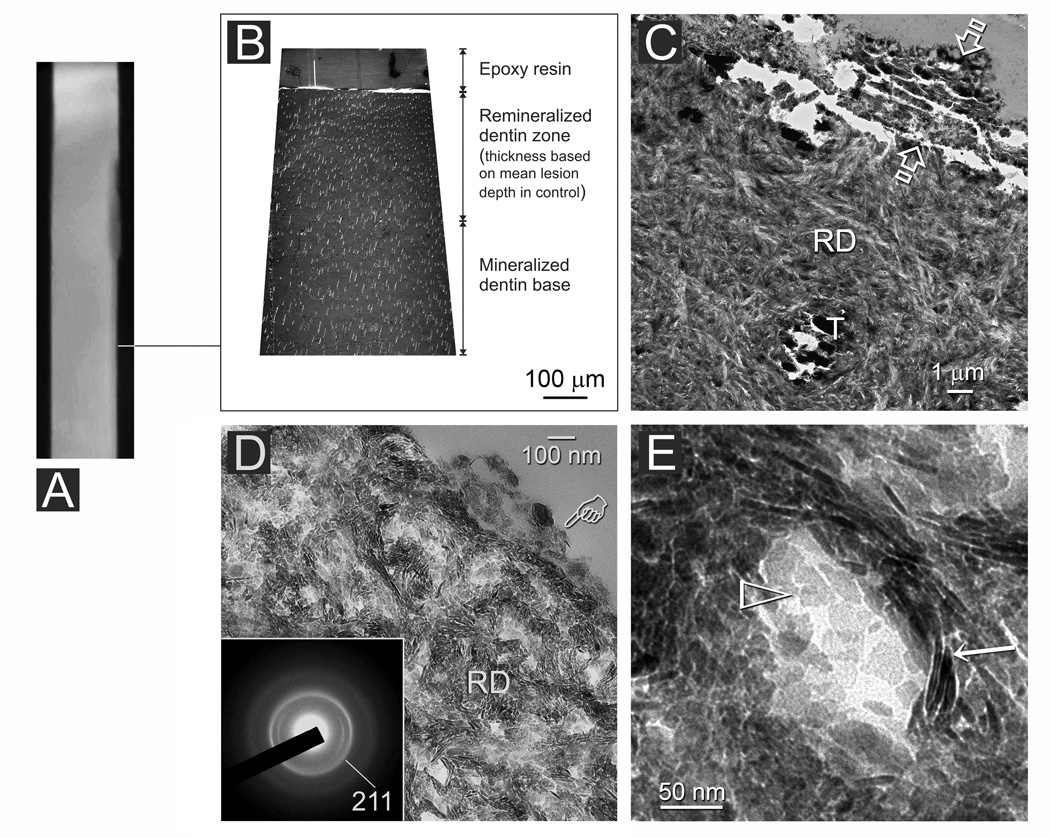
Fig. 4.
A non-bonded artificial caries-like dentin lesion after 4 months of biomimetic remineralization. A. A stacked 2-D micro-CT image indicating (line) the location from which TEM images were taken. B. Thick section of the remineralized lesion. At this magnification, there is no difference between the electron density of the remineralized dentin zone and the underlying mineralized dentin base. C. Thin section of the lesion surface with a layer of mineral precipitate (between open arrowheads). RD: Heavily remineralized dentin; T: dentinal tubule. D. Higher magnification of the remineralized dentin (RD) showing extrafibrillar and intrafibrillar remineralization. Pointer: surface mineral precipitates. Inset: selected area electron diffraction confirms the presence of apatite arranged along the longitudinal axis of the collagen fibrils. E. High magnification showing incomplete remineralization of the intrafibrillar space. Open arrowhead: intrafibrillar apatite platelets; Arrow: extrafibrillar apatite platelets.

