
Figure 2. Accumulation of protein-acrolein adducts and PF4 in the plasma and atherosclerotic lesions of acrolein-fed apoE-null mice.
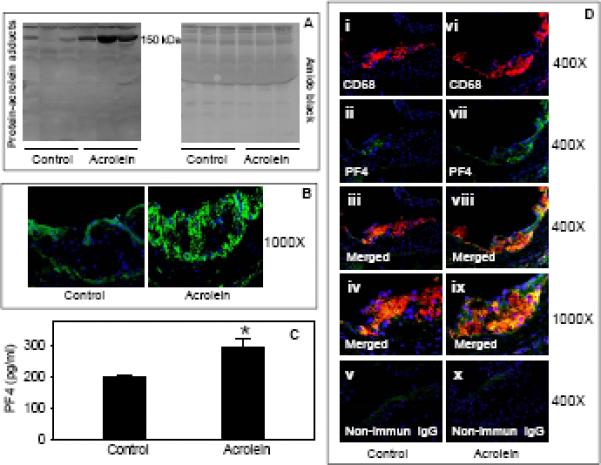
Eight week old male apoE-null mice were fed acrolein (2.5 mg/kg) or water (controls) by gavage for 8 weeks. A) Western blot analysis of protein-acrolein adducts in the plasma. Plasma obtained from control and acrolein-fed apoE-null mice, was probed with anti-acrolein-KLH antibody. B) Expression of protein-acrolein adducts in the aortic valve. Frozen sections of control and acrolein-fed apoE-null mice were stained with anti KLH-acrolein antibody, followed by Alexa488-conjugated goat-anti-rabbit secondary antibody. C) Plasma levels of PF4. Concentration of PF4 in the plasma was measured by sandwich ELISA. Values are means ± SEM. *P<0.01 vs controls. D) Expression and co-localization of PF4 with macrophages in the aortic valve. OCT-fixed frozen sections of control (i–v) and acrolein-fed (vi–x) apoE-null mice were stained with Alexa 647-conjugated anti-CD68 (i and vi; red) and PF4 (PF4; ii and vii; green, Alexa 488). Sections incubated with non-immune rabbit IgG (v and x) served as negative controls. The yellow fluorescence in the merged image (iii and viii) indicates PF4 co-localization with macrophages. Nuclei are identified in blue (DAPI). Panels iv and ix show higher magnification images of the colocalization of PF4 with macrophages.