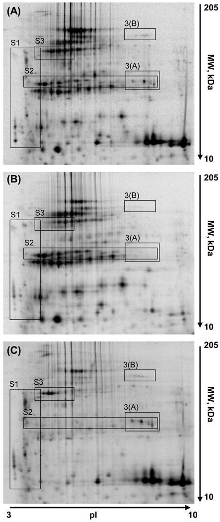
Figure 2.
2D-DIGE of total protein extracts from adipocytes and stromal vascular fraction cells isolated from abdomen adipose tissue. The figure presents the master gel image (A) and representative 2D-gel images from adipocytes (B) and stromal vascular fraction cell samples (C). An expanded view of the regions marked by rectangles are presented in Figures 3A, 3B and Supplementary Figures 1 – 3.

