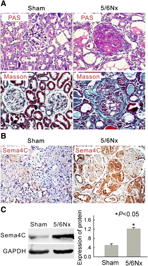
Fig. 1.
Expression of Sema4C is increased in the kidney tissue of 5/6 nephrectomized rats. (A) Representative histology of kidney sections from sham-operated and 5/6-nephrectomized (5/6 NX) rats stained with periodic acid–Schiff (upper) or Masson's trichrome stain (lower). Interstitial fibrosis and glomerular sclerosis are extensive in 5/6 NX rats. The tissue of sham rats was normal. Magnification × 200. (B) Representative immunohistochemical detection of Sema4C in the kidney tissue of sham-operated and 5/6-nephrectomized rats. Sema4C immunostaining is strong in the epithelial tubular cells in 5/6 NX rats and much weaker in sham rats. Magnification × 200. (C) Representative western blot of Sema4C in the kidney tissue of sham-operated and 5/6-nephrectomized rats. The expression of the Sema4C protein was elevated in the kidney tissue of 5/6-nephrectomized rats. The histogram shows the average volume density corrected for the loading control, GAPDH (n = 6). *P < 0.05 compared with sham-operated rats.

