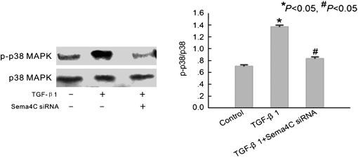
Fig. 3.
Depletion of Sema4C represses TGF-β1-induced activation of p38 MAPK. Representative western blot of phosphorylated p38 MAPK in control cells, TGF-β1-treated cells and TGF-β1-treated Sema4C-siRNA cells. The histogram shows the average volume density of phosphorylated p38 MAPK corrected for the loading control, total p38 MAPK (n = 4). *P < 0.05 compared with control cells. #P < 0.05 compared with TGF-β1-treated cells.

