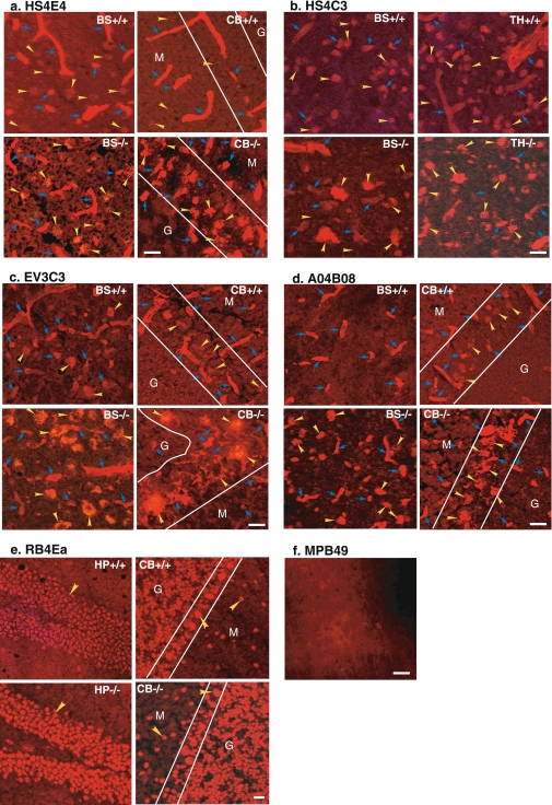
Fig. 1.
Differential distribution of HS domains in the brains of wt and MPS IIIB mice. Cryostat brain sections (10 μm) of 6-month-old wt and MPS IIIB mice were stained by immunofluorescence for specific HS domains using five antibodies targeting HS epitopes with different sulfation patterns: a HS4E4, b HS4C3, c EV3C3, d AO4B08, e RB4Ea. f MPB49 control. +/+ wt; -/- MPS IIIB, BS brain stem; CB cerebellum, TH thalamus, HP hippocampus. Yellow arrowheads: HS+ neurons. Blue arrows HS+ vasculature. G granular layer, M molecular layer; Purkinje cell layer (between two white lines). Scale bars: 20 μm

