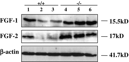
Fig. 2.
Increase in the expression of FGF1 and FGF2 in MPS IIIB mouse brain. Whole cell proteins (20 μg/sample) isolated from the brain of 6-month-old mice were subjected to SDS-PAGE separation and western blotting for FGF1, FGF2, or beta actin control. FGF2. +/+ 1–3: wt (n = 3); -/- 4–6: MPS IIIB (n = 3)

