
Fig. 1.
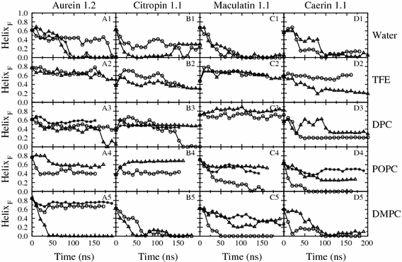
The helix fraction (HelixF) as a function of simulation time for the peptides aurein 1.2 (a), citropin 1.1 (b), maculatin 1.1 (c) and caerin 1.1 (d) in different environments. The environments were: water (1), 50% v/v trifluroethanol (TFE) (2), in the presence of a DPC micelle (3), in the presence of a POPC bilayer containing a pore (4) and in the presence of a planar DMPC bilayer (5). The triangles, circles and stars denote three independent simulations