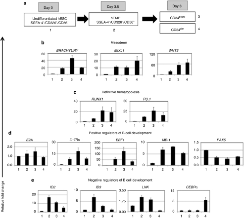
Figure 6.
Quantitative reverse transcription (RT)-PCR analysis of undifferentiated human embryonic stem cells (hESCs), early mesoderm progenitors (hEMP), and CD34bright and CD34dim cells. (a) Schematic model of stages of mesoderm and hematopoietic differentiation from hESCs under mesoderm-specific conditions [(bone morphogenetic protein-4 (BMP-4), basic fibroblast growth factor (bFGF), and vascular endothelial growth factor (VEGF) (no serum) (B+F+V)], showing immunophenotypic populations isolated for gene expression analysis by qRT-PCR. Day of isolation of each population is shown. Numbers 1–4 next to the boxes correspond to the populations analyzed by qRT-PCR in b–e. (b) Mesoderm-associated genes. (c) Genes associated with definitive hematopoietic development. (d) Positive regulators involved in B cell development and maturation. (e) Negative regulators that potentially block B cell development. Fold change in expression is shown on y-axis after normalization to housekeeping gene RPL-7. Mean ± SEM for three independent experiments using H9 cells.

