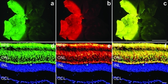
Figure 1.
Cotransduction of adeno-associated viruses (AAVs) in wild-type retinas. Eyes of wild-type mice were subretinally injected with a mixture of 1.5 × 10⁹ vector particles (vp) AAV-EGFP and 1.5 × 10⁹ vp AAV-DsRed. Two weeks postinjection eyes were fixed (n = 5), whole mounted for imaging, then cryosectioned (12 µm) and processed for histology. Nuclei were counterstained with DAPI. (a–c) Representative whole mounts illustrating (a) enhanced green fluorescent protein (EGFP), (b) DsRed, and (c) overlay of EGFP and DsRed signals. Representative sections demonstrate significant coexpression (f) of (d) EGFP and (e) DsRed signals at the cellular level in the outer nuclear layer. In order to obtain a clearer view of the markers the DAPI (blue) signal was edited out from the ONL. Bars = 1 mm (a–c) and 25 µm (d–f). GCL, ganglion cell layer; INL, inner nuclear layer; ONL, outer nuclear layer; RPE, retinal pigment epithelium.

