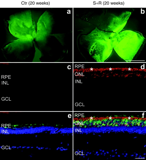
Figure 4.
Immunohistochemical analysis of rhodopsin expression following combined suppression and replacement therapy 20 weeks postinjection. The right eyes of P5 P347S mice were subretinally injected with a mixture of (b,d, and f) 6.0 × 10⁸ vp AAV-S and 1.8 × 10¹⁰ vp AAV-R whereas the left eyes were injected with (a,c, and e) 6.0 × 10⁸ vp AAV-C. Note that AAV-S and AAV-C coexpress enhanced green fluorescent protein (EGFP). Eyes were fixed (n = 5), whole mounted for imaging, then cryosectioned (12 µm) and processed for immunocytochemistry using rhodopsin primary and Cy3-conjugated secondary antibodies. Nuclei were counterstained with DAPI. (a,b) Representative whole mounts. (c,d) Representative sections show rhodopsin labeling (red). (e,f) Rhodopsin (red), EGFP (green), and nuclear DAPI (blue) signals overlaid. *Photoreceptor segment layer; ONL, outer nuclear layer; INL, inner nuclear layer; GCL, ganglion cell layer; RPE, retinal pigment epithelium. Bars = 1 mm (a,b) and 25 µm (c–f).

