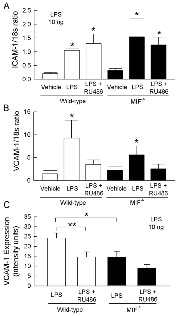
Figure 5. ICAM-1 and VCAM expression in wild-type and MIF−/− mice, and in the presence and absence of RU486.
A, B. Roles of endogenous MIF and GC in regulating LPS-induced expression of mRNA for ICAM-1 (A) and VCAM-1 (B) in muscle. Wild-type and MIF−/− mice were treated with vehicle alone, LPS (10 ng, 4 hrs) + vehicle, or with RU486 prior to LPS treatment. ICAM-1 & VCAM-1 mRNA were quantitated using real-time PCR and expressed relative to 18s mRNA. Data are shown as mean ± sem of n = 5–8. * denotes p<0.05 vs vehicle. C. Roles of endogenous MIF and GC in regulating LPS-induced VCAM-1 expression in the cremasteric microvasculature. Wild-type and MIF−/− mice were treated with LPS (10 ng) following pretreatment with RU486, or vehicle. Four hrs later, VCAM-1 expression was assessed using fluorescence microscopy to quantitate deposition of an Alexa 488-conjugated anti-VCAM-1 Ab. Data are shown for wild-type and MIF−/− mice following treatment with vehicle or RU486 (n=6/group). * p = 0.013 for MIF−/− vs wild-type vehicle-treated mice. ** p = 0.018 for effect of RU486 in LPS-treated wild-type mice.

