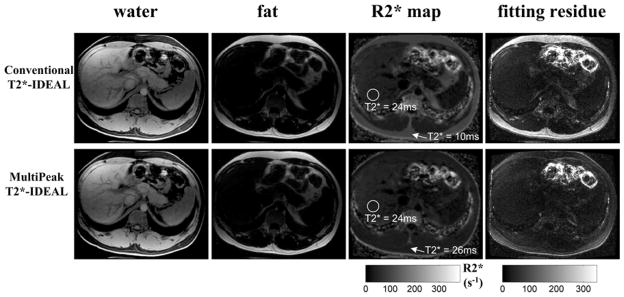
Figure 8.
Results from a 6-pt 3D-SPGR abdominal scan with a healthy volunteer. T2*-IDEAL reconstructions were performed with (second row) and without (first row) the multipeak correction (spectrum self-calibration). As can be seen from the R2* maps, conventional T2*-IDEAL results in an erroneous estimate of T2* in the subcutaneous fat (average T2* = 10ms). In contrast, the multipeak corrected R2* map shows an improved T2* estimate (averaged T2* = 26ms). The T2* values in liver remain the same as there is no fat in liver. As expected, the residue maps show significant improvement of the fitting in fatty tissues when using the multipeak model. Imaging parameters include: first TE = 1.1ms, echo spacing = 1.7ms, last echo = 9.5ms, TR = 16.2ms, 14 locations, BW = ±167kHz, FOV = 33cm×26cm, slice thickness = 8mm, matrix = 192×160, flip angle = 15 degrees and an 8-channel cardiac coil. The total imaging time was 30 seconds.

