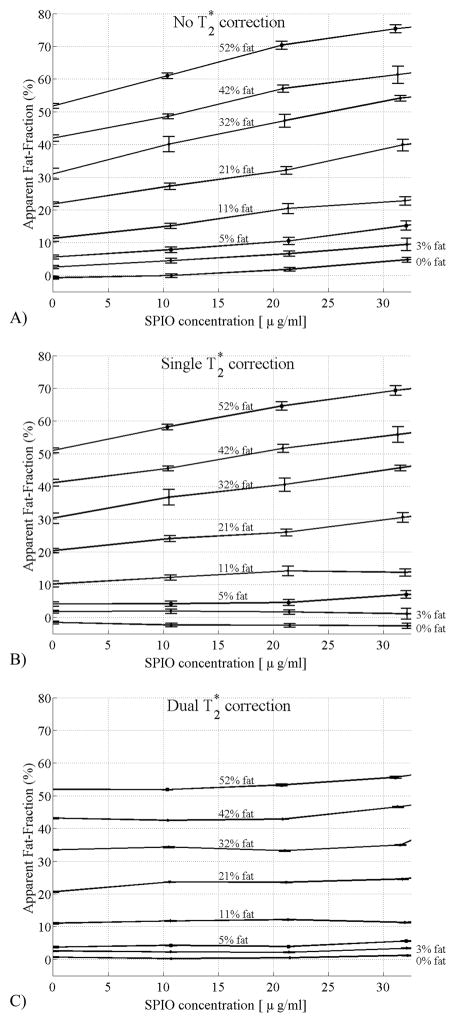
Figure 5.
Fat-fraction measured from the fat/water/SPIO phantom reconstructed with a) no T2* correction, b) single T2* correction, c) dual T2* reconstruction. Error bars show the standard error of the mean. As the SPIO concentration increases, the fat-fraction should remain constant if the correction algorithm is removing the effects of T2* decay correctly. Large errors are seen without T2* correction, and although the single T2* correction method improves estimates of fat-fraction, relatively large errors are still seen at high fat fractions. Only with the dual T2* correction method does the estimated fat-fraction agree closely with the known fat-fraction, independent of SPIO concentration.

